
Have you ever noticed dark specks drifting across your vision like tiny flies, or seen a brief flash of light at the edge of your eye when you turn your head in the dark? Many people dismiss these as eye strain or fatigue. But they can be early warning signs from the retina, and ignoring them can mean losing sight.
Retinal detachment is an eye emergency. Understanding its symptoms, risk factors, and treatment window can make the difference between preserved vision and permanent vision loss.
What Is Retinal Detachment?
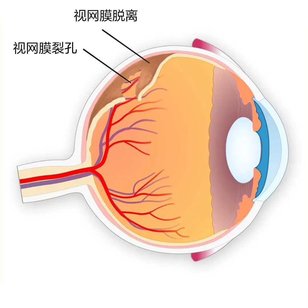
The retina is a thin, light-sensitive layer at the back of the eye, similar to the film in a camera. It captures light, converts it into visual signals, and sends those signals to the brain. When the retina separates from the underlying tissue, it is called retinal detachment.
Think of it like wallpaper peeling away from a wall. Once a small tear or hole forms, fluid can seep underneath and lift the retina off its normal position. When that happens, the affected area can no longer process images, causing vision loss.

The retina works like the light-sensitive film in a camera, converting images into signals for the brain.
Warning Signs Before Detachment
Retinal detachment rarely happens without warning. The eye usually sends signals first. Seek urgent ophthalmic evaluation if you experience any of the following symptoms.
Sudden increase in floaters: a shower of dark spots, threads, or cobweb-like shadows drifting in your vision.
Flashes of light: brief lightning-like streaks, especially in dim light or when moving the eyes.
Shadow or curtain in vision: a dark area that seems to spread from one side of your visual field.
Blurred central vision: the center of your vision becomes noticeably hazy.
Distorted vision: straight lines appear wavy or bent.

Common warning signs include floaters, light flashes, a curtain-like shadow, and blurred or distorted sight.
Who Is at Higher Risk?
Retinal detachment can affect anyone, but five groups have a significantly higher risk. Regular dilated eye examinations are especially important if you belong to one of these groups.
1. People with High Myopia
People with high myopia, generally 600 degrees or more, have longer eyeballs and thinner retinas. Like an over-stretched plastic film, the thinned retina is more likely to develop tears or holes. This is the largest high-risk group.
2. People with a History of Eye Trauma
Blunt impact or penetrating injury can create retinal breaks directly. Even injuries that seem minor can lead to retinal changes over time.
3. Older Adults
With age, the vitreous gel inside the eye naturally liquefies and shrinks. As it pulls away from the retina, it can tear the retinal surface.
4. People with a Family History
Genetic factors can influence retinal structure and vitreous behavior, increasing susceptibility to detachment.
5. People Who Have Had Cataract Surgery
Changes in the vitreous after cataract surgery can raise the risk of retinal tears or detachment, which is why postoperative follow-up is important.
Will Retinal Detachment Cause Blindness?
Retinal detachment is a medical emergency because it progresses quickly. Eye movement and gravity can enlarge the detached area. Once the macula, the central part of the retina responsible for sharp vision, is involved, central vision can be permanently affected.
The retina has a limited treatment window. Photoreceptor cells begin to die roughly 12 hours after detachment. For the best outcome, timing matters.
When the macula has not yet detached, prompt treatment usually leads to good vision recovery.
When the macula has detached, surgery within about one week still offers a reasonable chance, but final vision depends heavily on how long the macula was detached and how severe the detachment was.
A comprehensive dilated retinal exam allows the doctor to detect tears, holes, or early detachment before vision is permanently lost.
After Surgery: Four Keys to Recovery
Surgical success is only part of the outcome. Postoperative care strongly influences how well the retina heals and how much vision returns.
1. Maintain the Recommended Head Position
If gas or silicone oil is used to support the retina during surgery, the doctor will instruct you to keep a specific posture, often face-down or side-lying. This keeps the internal tamponade material pressing against the repaired area and helps the retina reattach. Failure to follow positioning instructions can lead to poor reattachment or recurrence.
2. Avoid Strenuous Activity
For one to three months after surgery, avoid rubbing the eyes, running, jumping, heavy lifting, and any activity that causes strong head movement or pressure changes in the eye.
3. Do Not Fly Until Cleared
If gas has been placed in the eye, air travel is unsafe until the gas has fully absorbed. Cabin pressure changes can cause dangerous rises in eye pressure. Always get clearance from your surgeon before booking a flight.
4. Attend All Follow-Up Visits
Regular follow-up allows the surgical team to monitor retinal reattachment, manage eye pressure, detect complications early, and plan removal of temporary filling materials when needed.
When to Seek Help
If you or someone you know suddenly develops flashes, a flood of floaters, or a curtain-like shadow in one eye, do not wait. Early evaluation and treatment by a retina specialist offer the best chance of preserving vision.
International patients can begin with a remote consultation to share symptoms and imaging, then arrange an in-person visit if urgent surgery is needed. Request a consultation →